George Town, Prayagraj, U.P.211002
Welcome to Dr. Aditi's Fetal Medicine Centre

Welcome to Dr. Aditi's Fetal Medicine Centre
George Town, Prayagraj, U.P.211002
Written by Dr. Aditi Dubey, M.B.B.S., M.S. (OBG), Fetal-Maternal Medicine Specialist, Dr. Aditi’s Fetal Medicine Centre, George Town, Prayagraj. More than 10 years of experience in fetal ultrasound and prenatal diagnosis.
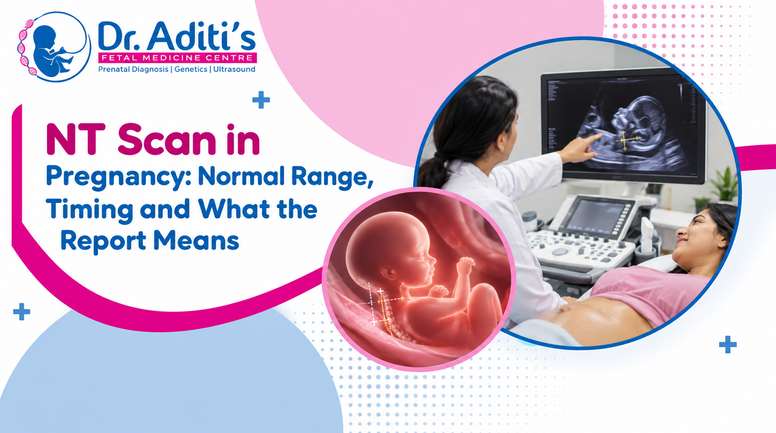
An NT scan in pregnancy is a first trimester ultrasound that measures the fluid layer at the back of your baby’s neck. It is done between 11 weeks 0 days and 13 weeks 6 days. An NT measurement below 3.0 mm is usually considered normal. A higher value means increased risk, not a diagnosis.
The NT scan in pregnancy is often the first detailed look a mother gets at her baby, and it is also the first real screening checkpoint of the pregnancy. Most of the worry around it comes from one thing, the report. This guide explains what the scan is, the exact week it should be booked, the normal NT range in mm, and how to read each line of your report without panic.
An NT scan in pregnancy is an ultrasound that measures the thin collection of fluid under the skin at the back of the baby’s neck. Every baby has this fluid layer in the first trimester. The measurement of that layer is called the nuchal translucency, or NT.
The thickness of this fluid is linked to the chance of chromosomal conditions such as Down syndrome, and to certain heart and structural problems. A thicker layer means a higher statistical chance. It does not mean the baby has a condition.
The NT scan in pregnancy is done through the abdomen with a normal ultrasound probe, exactly like any early pregnancy scan. It is painless and takes about 20 to 30 minutes, sometimes a little longer if the baby is lying in a difficult position.

The nuchal translucency scan full form is simply “nuchal translucency”, where nuchal means relating to the nape or back of the neck and translucency refers to how the fluid appears on ultrasound. Put together, an NT scan in pregnancy measures the clear fluid space behind the baby’s neck.
You may also see it written on your prescription as:
All of these refer to the same appointment at a fetal medicine centre in Prayagraj.
The NT scan in pregnancy is done in the third month, between 11 weeks 0 days and 13 weeks 6 days. In practice this means the scan is booked at any point from the start of week 11 to the last day of week 13.
The window is fixed for a technical reason. The measurement is only reliable when the baby’s crown rump length (CRL), which is the length from head to bottom, is between 45 mm and 84 mm. Before 11 weeks the baby is too small to measure accurately, and after 13 weeks 6 days the fluid layer starts to reabsorb and the reading loses its meaning.
Missing the NT scan in pregnancy window is the single most common problem we correct at the scan table. If your last menstrual period date is uncertain, book the appointment early rather than late. In our practice in Prayagraj we regularly see mothers arrive at 14 weeks and lose the chance to have the NT measured at all.
A normal NT scan in pregnancy measurement is generally below 3.0 mm, and most healthy babies measure between 1 mm and 2 mm. Because the neck fluid naturally increases as the baby grows, the normal range shifts slightly week by week, which is why your report compares your value against a chart rather than a single number.
The table below shows the NT scan in pregnancy normal range week by week, following the reference pattern maintained by the Fetal Medicine Foundation. Treat it as a guide to help you read your report, not as a verdict.

| Gestational age | Crown rump length (CRL) | Usual median NT | Approximate upper limit of normal |
|---|---|---|---|
| 11 weeks to 11 weeks 6 days | 45 to 58 mm | About 1.2 mm | Around 2.0 to 2.2 mm |
| 12 weeks to 12 weeks 6 days | 58 to 72 mm | About 1.4 mm | Around 2.3 to 2.5 mm |
| 13 weeks to 13 weeks 6 days | 72 to 84 mm | About 1.6 mm | Around 2.6 to 2.8 mm |
| Across the whole window | 45 to 84 mm | Under 2.0 mm in most pregnancies | 3.0 mm as a general cut off, 3.5 mm as clearly increased |
Two things matter more than the raw number in any NT scan in pregnancy report:
Along with the nuchal translucency, the first trimester scan checks several other markers. A complete NT scan in pregnancy report will usually include:
The scan is usually paired with a blood test on the same day. Together the ultrasound and the blood test form the combined first trimester screening. The blood component is the double marker test, which measures free beta hCG and PAPP-A.
Preparation for an NT scan in pregnancy is simple. Drink water and come with a moderately full bladder, because a full bladder pushes the uterus into a better position for the scan. Eat normally, there is no fasting requirement.
A high NT value on an NT scan in pregnancy report means the statistical chance of a chromosomal or structural condition is higher than average. It is a screening result, not a diagnosis. Most babies with an increased NT are born healthy, and the finding simply signals that a confirmatory test should be offered.
The next step depends on how high the reading is and what the combined risk works out to:
The national screening guidance published by the NHS follows the same principle, a screening result gives a chance, only a diagnostic test gives an answer.
Read the report in four steps: check the dates, check the NT number, check the markers, then read the final risk line. The risk line is the one that actually matters, and it is usually printed at the bottom.
| What the report says | What it means |
|---|---|
| NT = 1.5 mm | The nuchal fluid measurement, within the normal range |
| CRL = 62 mm | Baby’s length, confirms you are around 12 weeks 3 days |
| NB present | Nasal bone seen, a reassuring sign |
| FHR = 160 bpm | Fetal heart rate, normal for this stage |
| Risk 1 in 1500 | Low risk, roughly 1 pregnancy in 1500 with this profile is affected |
| Risk 1 in 120 | High risk band, further testing will be advised |
In screening, a bigger second number is better. A risk of 1 in 1500 is far more reassuring than a risk of 1 in 120. Most laboratories in India use a cut off around 1 in 250 to separate low risk from high risk.
The NT scan is done at 11 to 13 weeks 6 days and screens for chromosomal risk, while the TIFFA scan is done at 18 to 22 weeks and checks the baby’s organs and structure in detail. They are different scans at different stages, and one does not replace the other.
| Point of difference | NT scan | TIFFA / anomaly scan |
|---|---|---|
| Timing | 11 weeks 0 days to 13 weeks 6 days | 18 to 22 weeks |
| Main purpose | Chromosomal risk screening | Structural and organ assessment |
| Key measurement | Nuchal translucency in mm | Full organ by organ checklist |
| Blood test with it | Double marker, same day | Usually none |
| Duration | 20 to 30 minutes | 30 to 45 minutes |
| Can it be repeated later | No, the window closes | Yes, a repeat can be done if views are incomplete |
The NT scan in pregnancy is safe. It uses the same diagnostic ultrasound that has been used in pregnancy for decades, with no ionising radiation and no evidence of harm to the baby when performed by a trained operator for a medical reason.
It is strongly recommended but it is not legally compulsory. Skipping it means losing the earliest and most accurate window for chromosomal risk assessment, which is why most obstetricians advise it for every pregnancy, and not only for mothers above 35.
The accuracy of an NT scan in pregnancy depends almost entirely on who measures it and on how strictly the measurement protocol is followed. A difference of 0.3 mm can move a report from a reassuring risk figure to a high risk one, which is why the scan belongs with a trained fetal medicine specialist rather than a general sonography setup.

At our centre in George Town, Prayagraj, Dr. Aditi Dubey performs the first trimester scan personally, follows the standard measurement criteria for every case, and explains the report to the couple in the same sitting. A common situation we see is a mother arriving anxious about a borderline reading from an outside scan, where a correct repeat measurement within the valid window resolves the concern entirely. Full details of timing, inclusions and pricing are on the NT scan in Prayagraj page.
The NT window is short and it does not reopen. If you are between 10 and 13 weeks, book your NT scan in pregnancy appointment at Dr. Aditi’s Fetal Medicine Centre, George Town, Prayagraj, or call +91 83184 94849 to check the next available slot.
Statutory notice: This centre is registered under the PCPNDT Act 1994. Determination or disclosure of the sex of the foetus is not performed here and is not permitted by law, in any form or on any request.
Medical disclaimer: This article is for education only and is not a substitute for professional diagnosis or treatment. Please consult Dr. Aditi Dubey or a qualified fetal medicine specialist about your own pregnancy.
An NT scan in pregnancy is strongly recommended but it is not legally compulsory in India. It is the best early screening test for chromosomal risk, and it also confirms your dates and the number of babies. Most obstetricians advise it for every pregnancy, regardless of the mother’s age.
An abnormal NT means a higher statistical risk, not a confirmed condition. Your doctor will usually offer NIPT for further screening or amniocentesis for a definite answer, along with a detailed anomaly scan and a fetal echo later. Many babies with an increased NT are born completely healthy.
Yes, a moderately full bladder helps. Drinking two to three glasses of water about an hour before the appointment lifts the uterus into a better imaging position. You do not need to be uncomfortably full, and you do not need to fast before the scan.
No. The NT measurement alone cannot detect Down syndrome. It is combined with maternal age and the double marker blood test to calculate a risk figure. Confirmation requires a diagnostic test such as chorionic villus sampling or amniocentesis, which examines the baby’s chromosomes directly.
The NT cannot be measured after 13 weeks 6 days because the fluid layer reabsorbs. Screening then shifts to the quadruple marker test between 14 and 20 weeks, or to NIPT, which can be done from 10 weeks onwards at any later point in pregnancy.
The scan is painless and carries no known risk. It is performed on the abdomen with gel and a probe, with no needles and no radiation. The only discomfort most women report is from holding a full bladder for the duration of the appointment.