George Town, Prayagraj, U.P.211002
Welcome to Dr. Aditi's Fetal Medicine Centre

Welcome to Dr. Aditi's Fetal Medicine Centre
George Town, Prayagraj, U.P.211002
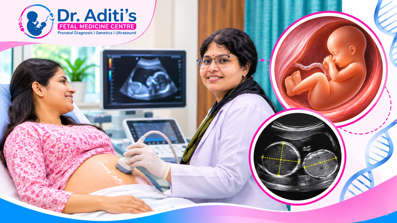
A growth scan in Prayagraj measures your baby’s head, abdomen, and thigh bone; estimates weight; and checks amniotic fluid and placenta. It is often performed during the third trimester, depending on clinical needs. Doctors may recommend it for suspected growth problems, twins, high blood pressure, diabetes, or previous pregnancy complications.
A fetal growth scan is a pregnancy ultrasound examination used to evaluate how a baby is developing inside the womb.
Unlike an early pregnancy scan, which confirms pregnancy location and viability, a growth scan focuses on fetal measurements, estimated weight, amniotic fluid, and relevant placental findings.
At Dr. Aditi’s Fetal Medicine Centre, Dr. Aditi Dubey assesses fetal measurements alongside gestational age, maternal health history, and previous ultrasound reports.
| Measurement | What It Tells the Doctor |
|---|---|
| Biparietal Diameter (BPD) | Measures the width of the baby’s head |
| Head Circumference (HC) | Helps assess head growth |
| Abdominal Circumference (AC) | Helps evaluate fetal growth and nutritional status |
| Femur Length (FL) | Measures the baby’s thigh bone |
| Estimated Fetal Weight (EFW) | Calculates approximate fetal weight using ultrasound measurements |
| Amniotic Fluid Index (AFI) | Helps assess the amount of amniotic fluid |
| Placental Assessment | Evaluates placental location and relevant visible features |
Doctors compare these measurements with reference charts for the baby’s gestational age.
A single measurement outside the expected range does not always indicate a problem. The doctor considers growth percentiles, previous scans, and other clinical findings.
Learn more about 2D ultrasound and its role in pregnancy assessment.
A fetal growth scan is often recommended during the third trimester, commonly around 28–36 weeks when clinically indicated.
However, the exact timing depends on the pregnancy and the reason for the examination.
If the mother’s abdominal measurements suggest that the baby may be smaller than expected, an ultrasound can help assess fetal size.
The doctor may investigate whether the baby is naturally small or whether fetal growth restriction is suspected.
Some babies measure larger than average for their gestational age.
A growth scan can estimate fetal weight and help doctors evaluate possible contributing factors, including maternal diabetes.
Maternal hypertension and preeclampsia may affect placental function and fetal growth.
Additional ultrasound monitoring may be recommended to assess the baby’s development.
Gestational diabetes and pre-existing diabetes can influence fetal growth.
Your doctor may recommend additional monitoring to identify unusually rapid or reduced growth.
Twin pregnancies often require more frequent ultrasound assessments.
The recommended schedule depends on whether the babies share a placenta and whether any complications are present.
Women with a history of fetal growth restriction, stillbirth or certain placental complications may need closer monitoring.
Patients with additional pregnancy risk factors can consult a high risk pregnancy specialist.
During a growth scan in Prayagraj, fetal measurements are compared with expected values for the pregnancy week.
The findings are commonly expressed as percentiles, which help doctors understand how the baby’s measurements compare with reference populations.
A baby may be described as small for gestational age when estimated fetal weight or abdominal circumference is below the 10th percentile.
However, a smaller measurement does not automatically indicate a health problem.
Some babies are naturally smaller because of constitutional or genetic factors.
Doctors consider additional clinical findings before determining whether further investigation is required.
Fetal growth restriction means the fetus has not reached its expected growth potential.
Possible contributing factors include:
When fetal growth restriction is suspected, doctors may recommend Doppler ultrasound, repeat measurements, and closer monitoring.
A baby may be described as large for gestational age when estimated fetal weight exceeds the 90th percentile.
Maternal diabetes, genetic factors, and previous pregnancy history may contribute.
Important: Ultrasound provides an estimated fetal weight, not an exact birth weight. Doctors interpret the estimate alongside other pregnancy findings.
A growth scan evaluates more than fetal size.
Amniotic fluid and placental findings are important components of pregnancy assessment.
Amniotic fluid surrounds the developing baby and supports normal development.
Doctors may measure fluid using the Amniotic Fluid Index (AFI) or Single Deepest Vertical Pocket (SDVP).
These measurements can help identify:
Abnormal fluid findings may require further assessment depending on gestational age, maternal health and fetal wellbeing.
The placenta supplies oxygen and nutrients to the developing baby.
During ultrasound, the doctor may assess placental location and other relevant features.
If there is concern about placental function or fetal growth, Doppler ultrasound may be advised.
Doppler evaluates blood-flow patterns in selected blood vessels, including the umbilical artery.
Read more about the Doppler scan in Prayagraj and how it supports the monitoring of certain high-risk pregnancies.
The cost of a growth scan in Prayagraj depends on the type of examination, whether additional assessments are required and the centre’s billing policy.
A routine fetal growth assessment and a detailed examination with Doppler may have different charges.
| Cost Factor | Explanation |
|---|---|
| Type of ultrasound | Routine growth assessment or detailed evaluation |
| Doppler examination | Additional blood-flow measurements may affect charges |
| Multiple pregnancy | Twin pregnancies may require additional measurements |
| Specialist consultation | Consultation charges depend on the centre’s policy |
| Follow-up examination | Repeat ultrasound examinations may have separate charges |
Before booking, patients should confirm whether the quoted amount includes the written ultrasound report, specialist consultation and Doppler assessment if required.
The examination should be selected according to medical requirements rather than cost alone.
Dr. Aditi Dubey is a fetal medicine specialist with a Postgraduate Degree in Fetal Cardiology from Fetal Medicine Barcelona.
Dr. Aditi’s Fetal Medicine Centre is located in George Town, Prayagraj.
A growth scan in Prayagraj at the centre is intended to help assess fetal development, estimated weight, amniotic fluid and relevant pregnancy findings.
Patients should bring their previous ultrasound reports and pregnancy records so the doctor can review changes over time.
Ultrasound estimates fetal weight using measurements such as head circumference, abdominal circumference and femur length. The estimate commonly has an error margin of around 10-15%, although errors can be larger. Doctors therefore interpret estimated weight alongside growth trends, gestational age and other clinical findings rather than treating it as exact.
The number of growth scans depends on your pregnancy history and medical condition. Some pregnancies need only one third-trimester assessment, while high-risk pregnancies may require repeated monitoring. When evaluating growth trends, scans are generally spaced sufficiently apart, often around three weeks, unless another clinical reason requires earlier ultrasound assessment.
No. A growth scan measures fetal size, estimated weight, amniotic fluid and relevant placental findings. Doppler ultrasound evaluates blood-flow patterns in selected blood vessels. Your doctor may recommend both examinations when monitoring fetal growth restriction, hypertension or other pregnancy complications. Doppler is not automatically necessary for every pregnancy.
If your baby appears to be growing slowly, your doctor may recommend repeat ultrasound measurements, Doppler assessment and closer pregnancy monitoring. Management depends on gestational age, growth trends and fetal wellbeing. Patients seeking a fetal growth scan near george town prayagraj should bring previous ultrasound reports for comparison.